Multi-photon microscopy, also known as 2-photon microscopy or two-photon microscopy, is a widely used technique in the biological sciences, where sum-frequency generation within the sample can result in second harmonic generation or third harmonic generation to excite fluorescence in the sample. This process is widely used for creating fluorescence images of live cells, since the multiphoton microscopy lasers are typically in the near-infrared wavelengths region, therefore reducing the potential of photochemical damage to the cells. Due to the non-linear nature of this process, multiphoton microscopy lasers must have extremely short pulses, and therefore mode-locked femtosecond lasers are typically used for this application. Additionally, as in all microscopy applications, multiphoton microscopy lasers, must also be single spatial mode to get diffraction-limited performance out of the microscope objective. On this page you will find a list of our mode-locked multiphoton microscopy lasers, these mode-locked fiber lasers are ideal for multiphoton microscopy because of their short pulse width and excellent beam quality.
Multi-photon microscopy is utilized for numerous life science applications throughout the fields of physiology, neurobiology, embryology, and tissue engineering. Near-transparent tissues (e.g., skin cells) can be easily visualized in great detail thanks to this microscopy method. Noninvasive optical biopsy procedures benefit from two-photon microscopy’s high-speed imaging capabilities. Localized chemical reactions can be produced when working within cell biology. 2-photon microscopy is also beneficial for cancer research (characterizing skin cancer, revealing tumor cell arrest, tumor cell-platelet interaction, etc.), in the neurosciences (characterizing intact neural tissues), and in brain in-vivo imaging. In-vivo imaging of the brain benefits from this process because of the ability for deep imaging in scattering tissue. Two-photon microscopy is often used to image the live-firing of neurons in model organisms, such as fruit flies, mice, and zebrafish.
Lasers and Multi-Photon Microscopy
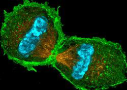
Fluorescence microscopy is deployed widely in biological sciences for identifying the spatial distribution of molecules of interest in complex heterogeneous samples, including living cells. Over the past 100 years, this method has been heavily dependent on the use of fluorescent tags which have uniquely engineered excitation and absorption spectra, and are functionalized to bond with particular molecules of interest. Not only do these tags increase the technique’s specificity, but they also reduce photochemical degradation of the sample by moving from ultra-violet to visible excitation wavelengths. For many years the advantages of fluorescence tags outweighed the disadvantages of the sample preparation requirements, but with the emergence of two-photon fluorescence microscopy, it is now possible to achieve many of these same advantages without any sample pretreatment. As a result, two-photon microscopy, also referred to as multiphoton microscopy, has once again revolutionized the field of biological imaging allowing for the creation of high-resolution images of living cells.
Multiphoton absorption as an excitation source for fluorescence was first theorized as far back as the early 1930s, 30 years before the invention of the laser enabled the construction of the two-photon microscope at Cornell University. But it is important to note that it wasn’t just any laser that would facilitate two-photon absorption; this process is only achieved when utilizing an ultrafast mode-locked laser. To understand why this application requires ultra-fast laser pulses, you must first recall that the sum frequency generation, also referred to as second-harmonic generation, is a nonlinear process. Therefore, this process is dependent on the intensity of the lasers electric field; hence, a tighter focus and a shorter pulse width will result in more efficient two-photon absorption. For a diffraction-limited microscope, the spot size at the sample is determined solely by the numerical aperture of the objective lens and the laser wavelength, that leaves the pulse duration as the only significant variable in the system. For this reason, mode-locked lasers which produce ultra-short pulses (on the order of 100 fs) at high repetition rates have become the standard excitation source for two-photon microscopy.
While the first two-photon system was demonstrated using a mode-locked dye laser, the apparent drawbacks associated with dye laser technology prevented any sort of commercialization at the time. It wasn’t until the advent of the Ti:Sapphire laser that such systems started to evolve from a physics demonstration, into a true analytical instrument. The Ti:Sapphire was a vast improvement over other early mode-locked laser sources, but it was far from an ideal solution. One of the biggest reasons for this fact is that the majority of fluorescent proteins of interest to biologists have a two-photon absorption band primarily in the 900 nm to 1100 nm range, while the peak lasing efficiency of Ti:Sapphire is around 780 nm. Luckily now there is a wide variety of lower-cost, fit for purpose mode-locked lasers sources available on the market.
How Mode-Locked Lasers Affect the Past, Present, and Future of Two-Photon Microscopy
In Winfried Denk’s original work he utilized a mode-locked dye laser to produce approximately 100 fs pulses at 630 nm to excite a cluster of 9 m fluorescent latex beads. While dye lasers may be suitable for demonstration in a university laboratory, they are far from commercializable. As a result the standard expiation source for two-photon microscopes quickly became the model-locked Ti:Sapphire laser. Aside from the obvious advantages of utilizing a solid state laser source, the Ti:Sapphire was also advantageous because it is widely tunable in the near infrared region, which causes minimal damage to samples and can penetrate much deeper into these samples compared to other commonly used visible wavelengths.
While Ti:Sapphire represented a huge improvement over other early mode-locked laser sources, it does have one major drawback. The peak lasing efficiency of Ti:Sapphire is around 780 nm while the majority of fluorescent proteins of interest to biologists have a two-photon absorption band primarily in the 900 nm to 1100 nm range, as shown in Figure 2 above. Additionally, it has been shown that there is a significant decrease in damage to living tissue by tuning the laser from 780 nm to 920 nm [2]. As a result, in most two-photon microscopes the laser is tuned to 920 nm where unfortunately Ti:Sapphire lasers are far less efficient.
Higher Power fs Fiber Lasers to Image Better, Deeper & Faster
Recent technological developments led to the commercial availability of new generations of femtosecond lasers capable of offering a wider variety of pulse regimes. Femtosecond lasers based on Optical Parametric Chirped-Pulse Amplifications (OPCPA) offer the widest range of pulse regimes, capable of operating at low and selectable repetition rates, in the µJ level and covering a wide wavelength range from near infrared to infrared wavelengths. Although OPCPA based systems offer high versatility, they are bulky, expensive, not as reliable and stable as conventional lasers.
Alternatively fixed wavelength femtosecond lasers have proved to offer a good compromise for 2-photon excitation microscopy. Their main advantages reside in its low price, very compact and robust format, high electrical efficiency and high reliability. This novel generation of lasers available at 780 nm, 920 nm and in the 1030-1070 nm wavelength range were initially conceived to match standard pulse regimes of Ti:Sapphire lasers to replace or complete this solution. Fixed wavelength femtosecond lasers are now offering alternative pulse regimes not accessible by Ti:Sapphire lasers such as various repetition rates, higher average power and higher energy making them ideal candidates to push boundaries of today’s research. Nevertheless, with such a wide variety of femtosecond lasers, choosing an adequate laser solution suitable for a given application is becoming challenging.
Two-Photon Microscopy at the Harvey Lab – Harvard Medical School
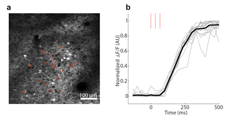
In Dr. Harvey’s particular case the presence of the ChrimsonR actuator was verified using two-photon fluorescence microscopy with an excitation wavelength of 1060nm. The figures below show the in vivo, two-photon calcium imaging performed with the DIADEM-IR-30. Figure (a) shows an example of a two-photon field of view in mouse parietal cortex where the individual neurons were sequentially stimulated with 3 x 30 ms spiral scans where the red asterisks indicate stimulation sites. Figure (b) shows the normalized ΔF/F responses of target neurons, which represents the normalized change in fluorescence intensity over time showing the rate of excretion on the ChrimsonR optogenetic actuator over time. The data from these two figures show how Dr. Harvey was able to use two-photon microscopy to verify the success of genetic modifications.
Additionally, his group also provided us with the video below showing the release of the ChrimsonR actuator in real time. To produce this video, he used a spatial light modulator to generate beamlets targeted to each neuron indicated by a red arrow, then simultaneously activated them using 10mW of average power per cell. As the stimulation laser activates the cells, their fluorescence increases as a result of calcium entering the cell and binding the calcium indicator. As in the previous example, the stimulation was done using the DIADEM -IR-30 mode-locked laser from Spark Lasers, which was customized to lase at 1060nm.
The New Alcor 1064nm 5W Femtosecond Laser for Multi-Photon Microscopy
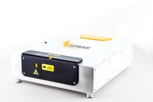
This 5W 1064nm version provides excellent pulse characteristics, offering >62nJ of pulse energy at a fixed repetition rate of 80MHz (40MHz also available), with a pulse duration of <130fs, for a peak pulse power of >480kW! The extremely high peak power of the ALCOR 1064-5W provides better brightness and contrast for your 2-photon microscopy application. This version comes standard with GDD precompensation from 0 down to -30,000fs^2 (others optional), conveniently tunable from the laser’s user interface. With negative GDD precompensation applied in the laser head, pulse duration is kept the same from laser output to beam excitation on the cells/tissues, despite any optics that would induce dispersion on the pulse. The ALCOR series also provides a ‘clean pulse’ by minimizing side lobes to avoid any thermal effect around the treated cell. SPARK Lasers specifically designed and developed the ALCOR 1064-5W for larger surface imaging for multi-photon microscopy, allowing a higher level of brilliance and contrast for two-photon excitation of calcium indicators such as GCaMP of other red opsins (RCaMP)
2-Photon Microscopy: XSight’s Vision for a Simplified Experience
In two previous blogs, we discussed the capabilities of the ALCOR Femtosecond Laser, successful applications, and briefly outlined two optional, external modules: the XSight and FLeX Fiber modules for further enhancing the capabilities of the various ALCOR configurations. In this article, we’re going to focus a bit more on the XSight Module, and how this addition can help your project succeed.
The ALCOR Series of femtosecond lasers can be equipped with the optional XSight, fully-integrated electronics module for 920nm & 1064nm, allowing for easy power adjustments and modulation. By itself, the ALCOR is an excellent laser for Two-Photon Imaging applications, offering fixed output powers of 1W, 2W, 4W, and 5W, but has no power modulation, and can only operate in alignment power mode, or full power mode, with no intermediate power levels. With the addition of the XSight module, users can tailor the output power to achieve the best results from their specific application. Rather than offering one configuration with all the bells and whistles, the modular design of the ALCOR Series allows you to save money on unnecessary components if your project doesn’t require the advanced features. Additionally, while there is a loss of total transmitted power when adding the XSight module, the transmission efficiency is one of the highest on the market for this type of laser, coming in at an impressive 85%.
Context-Dependent Sensory Processing Across Primary and Secondary Somatosensory Cortex
Are you looking for a high-power, high repetition rate, femtosecond laser for your bioimaging application? Check out our newest white paper, titled “Context-Dependent Sensory Processing Across Primary and Secondary Somatosensory Cortex.” This white paper, provided by Jerry Chen of Chen Labs at Boston University, details their successful experiment, investigating the roles of the primary and secondary somatosensory cortex during a tactile working memory task in mice. Within the experiment, in vivo calcium imaging of RCaMP1.07 imaging was performed using a 40MHz, 1040nm, 5W Altair fiber laser from SPARK Lasers.
jYCaMP: An Optimized Calcium Indicator for Two-Photon Imaging at Fiber Laser Wavelengths
Check out our newest white paper, titled “jYCaMP: An Optimized Calcium Indicator for Two-Photon Imaging at Fiber Laser Wavelengths.” This white paper, provided by Jerry Chen of Chen Labs at Boston University, details their successful experiment, identifying spectral variants of the recently developed jGCaMP7 family of GECIs, resulting in the development of jYCaMP, a red-shifted variant, perfectly suited for fiber laser wavelengths. Within the experiment, Chen Labs utilized a SPARK Lasers Altair, 31.25MHz, 1040nm, femtosecond fiber laser.
Large Volume Two-Photon Neural Imaging, Maintaining Cellular Resolution
Read about large volume two-photon imaging with cellular resolution in this white paper, titled “Flexible Simultaneous Mesoscale Two-Photon Imaging of Neural Activity at High Speeds,” provided by Jerry Chen of Chen Labs at Boston University and his colleagues.
Abstract: Understanding brain function requires monitoring local and global brain dynamics. Two-photon imaging of the brain across mesoscopic scales has presented trade-offs between imaging area and acquisition speed. We describe a flexible cellular resolution two-photon microscope capable of simultaneous video rate acquisition of four independently targetable brain regions spanning an approximate five-millimeter field of view. With this system, we demonstrate the ability to measure calcium activity across mouse sensorimotor cortex at behaviorally relevant timescales.
With many years experience providing multi-photon microscopy lasers to researchers and 100s of units fielded, we have the experience to ensure you get the right product for the application. Working with RPMC ensures you are getting trusted advice from our knowledgeable and technical staff on a wide range of laser products. RPMC and our manufacturers are willing and able to provide custom solutions for your unique application.
If you have any questions, or if you would like some assistance please Contact Us here. Furthermore, you can email us at info@rpmcdev.maxdroplet4.maxburst.devto talk to a knowledgeable Product Manager.
Alternatively, use the filters on this page to assist in narrowing down the selection of multi-photon microscopy lasers for sale. Finally, head to our Knowledge Center with our Lasers 101 page and Blogs, Whitepapers, and FAQ pages for further, in-depth reading.
Finally, check out our Limited Supply – In Stock – Buy Now page: This page contains an ever-changing assortment of various types of new lasers at marked-down/discount prices.