Traditional microscopy is the technical field of using microscopes to view objects and areas (mostly cells) that are not within the resolution range of the naked eye. Compound microscope designs feature objectives and condensers, but also consists of very simple, single-lens instruments that are often hand-held, such as a photography loupe or common magnifying glass.
Optical microscopy and electron microscopy involve the diffraction, reflection, or refraction of electromagnetic radiation/electron beams interacting with the specimen, and the collection of the scattered radiation or another signal in order to create an image. This process may be carried out by wide-field irradiation of the sample (for example standard light microscopy and transmission electron microscopy). This technique, though useful, has a few drawbacks: it only allows for scanning the surface of tissues, offers relatively low resolution and contrast, and is one-dimensional.
|
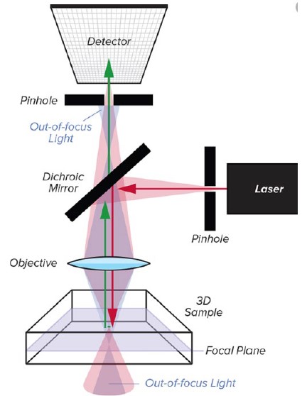
Confocal microscopy, or confocal laser scanning microscopy (CLSM) is an optical imaging technique for increasing optical resolution and contrast of a micrograph by using a spatial pinhole to block out-of-focus light in image formation. This technique is used extensively in the scientific and industrial communities, and typical applications are in life sciences, semiconductor inspection and materials science. Light travels through the sample under a conventional microscope as far into the specimen as it can penetrate, while a confocal microscope only focuses a smaller beam of light at one narrow depth level at a time. The CLSM achieves a controlled and highly limited depth of focus. A couple benefits of this technique include the ability to generate multiple two-dimensional images at various depths,and also enables the reconstruction of three-dimensional structures (i.e. optical sectioning) within an object. However, it can cause a photobleaching effect in the sample, can be adversely affected by background light, and due to its small field of view (FoV) can be a relatively slow process, compared to other techniques. | 
|
Pros: |
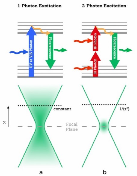
Two-photon excitation microscopy (TPEF or 2PEF) is a fluorescence imaging technique that allows imaging of living tissue up to about one millimeter in thickness. Unlike traditional fluorescence microscopy, in which the excitation wavelength is shorter than the emission wavelength, two-photon excitation microscopy typically uses near-infrared (NIR) excitation light which can also excite fluorescent dyes. However, for each excitation, two photons of NIR light are absorbed. Using infrared light minimizes scattering in the tissue. Due to the multi-photon absorption, the background signal is strongly suppressed.
|
 |
|
Cons:
|
|
Ti-Sapphire lasers are:
|
Femtosecond fiber lasers are:
|
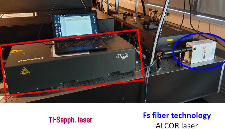
The fiber based design of the Alcor laser system enables a more compact, robust, and reliable laser than its DPSS counterparts, while still being air-cooled. Through this simplified fiber design, the Alcor requires little to no maintenance. Historically, researchers have been using Ti: Sapphire lasers, which utilize many more components and moving parts, requiring significantly more maintenance and ultimately leading to higher total cost of ownership. In the Alcor, the only consumables are the laser diodes, which are rated for >20,000 hours.
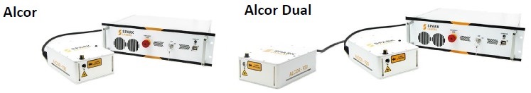
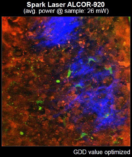
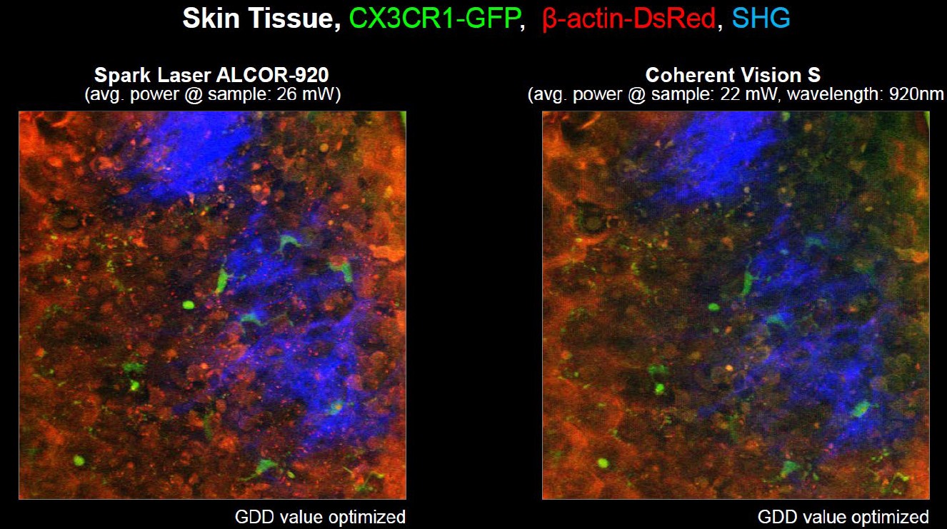
The Alcor femtosecond laser series features the most output power commercially available today, offering versions with up to 5W at 1064nm, and up to 4W at 920nm. The Alcor Dual is available with up to 2W of power with both 920nm and 1064nm outputs. With a <140fs pulse at 80MHz (others optional), the Alcor series is specifically designed for biophotonics applications such as multiphoton microscopy. The combination of ultra short pulse duration, high repetition rates and high average powers offers many benefits to researchers and OEM microscopy instrumentation manufacturers in the lifesciences and biophotonics fields. With up to 50 nJ of pulse energy and >384 kW of peak power, the Alcor enables higher brilliance and contrast in two-photon imaging of green fluorophores (GFP) and calcium indicators such as GCaMP.
In addition, the ALCOR can be equipped with the XSight (AOM) module for precise and fast power control, as well as the FleX fiber-coupled output module.
XSight – with fine and fast power control :
|
FLeX Fiber – fiber delivered fs pulses with total pulse control :
|
The addition of the Alcor FLeX fiber module allows you to turn your traditional confocal microscope in to a Two-Photon Microscopy scope, by injecting femtosecond pulses directly into any confocal microscope via the 2m long fiber optic cable!
Be sure to look out for the follow-up to this article, in which we will take a deeper dive into the Alcor series and some of the newest advancements, its XSight and FLeX fiber modules, technical specifications and successful experiments and applications using the Alcor series and other lasers from SPARK Lasers!
To get more information about any of the Alcor configurations, check out SPARK Lasers‘ page, or talk to one of our knowledgeable Product Managers today, by calling 1.636.272.7227 or click the link below to contact us!
Talk to a Product Manager

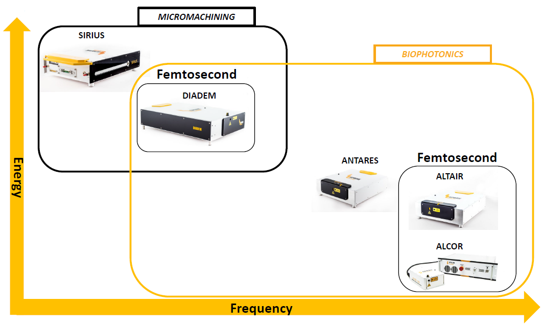
SPARK Lasers are experts in designing and manufacturing cutting-edge, reliable, and user-friendly lasers, offering several diverse product families, with a range of energies and frequencies and geared toward a variety of micromachining and biophotonics applications. SPARK’s product families include the Alcor, Altair, Antares, Diadem, and Sirius series. |
 |